Diagnosis and treatment of corneal diseases
The cornea is the clear, dome-shaped tissue that covers the front of the eye, playing a vital role in focusing light onto the retina. Corneal diseases can disrupt vision and affect the eye’s overall health. Early diagnosis and treatment of corneal diseases are essential to preserve sight and prevent complications.
This comprehensive guide will explore how corneal diseases are diagnosed and the most effective treatment options available.
What are Corneal Diseases?
Corneal diseases encompass a variety of conditions that affect the cornea’s structure, transparency, or function. They can range from minor injuries to complex medical conditions, such as infections, genetic disorders, or degenerative conditions.
Common Types of Corneal Diseases
Corneal Dystrophies:
- Genetic conditions leading to the accumulation of abnormal deposits in the cornea.
- Examples: Fuchs’ endothelial dystrophy, lattice dystrophy, and granular dystrophy.
Corneal Infections:
- Caused by bacteria, fungi, viruses, or parasites.
- Examples: Bacterial keratitis, fungal keratitis, viral keratitis (e.g., herpes simplex).
Corneal Ulcers:
- Open sores on the cornea, often resulting from untreated infections or trauma.
Keratoconus:
- A condition in which the cornea thins and bulges into a cone-like shape, impairing vision.
Dry Eye Syndrome:
- Inadequate tear production leads to inflammation and irritation of the cornea.
Trauma or Injury:
- Physical injuries such as scratches, foreign bodies, or burns can damage the cornea.
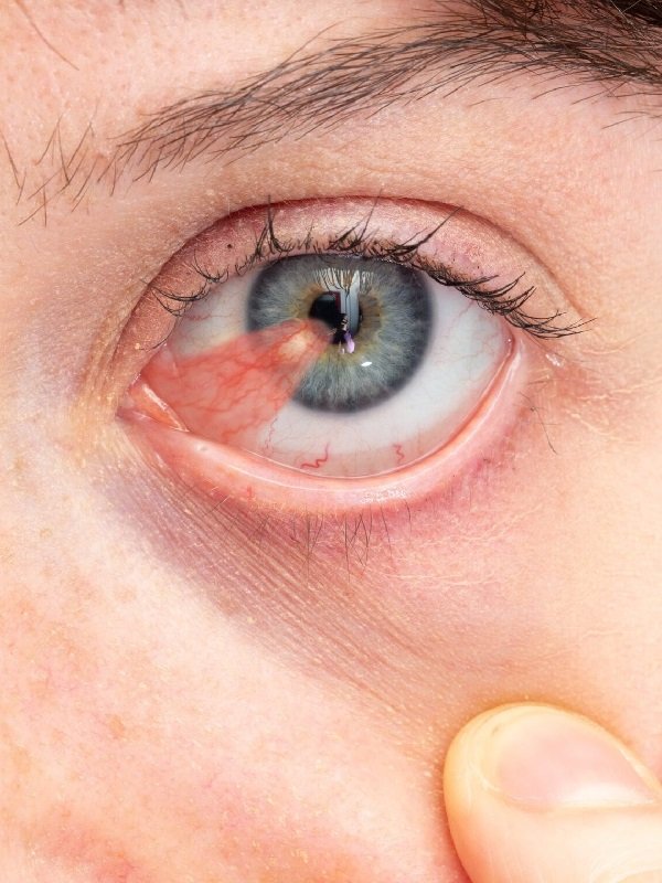
Pterygium:
- A growth of tissue from the conjunctiva onto the cornea, often related to UV exposure.
Understanding these conditions is key to early intervention and treatment.
Diagnostic Tests for Corneal Diseases
Slit-Lamp Examination:
- A microscope used to examine the cornea and other structures of the eye in detail.
- Detects signs of injury, disease, or structural changes.
Corneal Topography:
- A mapping technique that creates a 3D image of the cornea to detect irregularities or shape changes, as seen in keratoconus or post-surgical conditions.
Pachymetry:
- Measures the thickness of the cornea, which can be affected by diseases like keratoconus or Fuchs’ dystrophy.
Anterior Segment Imaging:
- High-resolution imaging, such as optical coherence tomography (OCT), provides cross-sectional views of the cornea and anterior eye structures.
Fluorescein Staining:
- A dye is applied to the eye to identify corneal ulcers, abrasions, or areas of dryness.
Culture Tests:
- When infections are suspected, a sample of corneal tissue or fluid may be cultured to identify bacteria, fungi, or viruses.
Tear Break-Up Time (TBUT):
- Tests for dry eye by measuring the stability of the tear film on the cornea.
Treatment Options for Corneal Diseases
The treatment of corneal diseases depends on the underlying cause, severity, and specific condition. Treatments may range from medication to surgical interventions, depending on the diagnosis.
1. Medical Treatments
a) Antibiotic or Antifungal Drops for Infections
- Bacterial Keratitis: Treated with topical antibiotics to eliminate bacterial infections.
- Fungal Keratitis: Requires topical antifungal medication.
- Viral Keratitis: Often treated with antiviral eye drops or systemic antiviral medication, depending on the severity.
b) Anti-inflammatory Eye Drops
Used for conditions like Fuchs’ dystrophy or severe dry eye to reduce inflammation and promote healing.
c) Artificial Tears for Dry Eye Syndrome
Lubricating eye drops or prescription eye drops can help alleviate symptoms and improve corneal health.
2. Surgical Treatments
a) Corneal Transplant (Keratoplasty)
- Full-Thickness Corneal Transplant: Replaces the entire cornea with a donor cornea.
- Partial-Thickness Corneal Transplant (Lamellar Keratoplasty): Replaces only part of the cornea, preserving more of the patient’s own corneal tissue.
- Common for advanced corneal scarring, keratoconus, or severe infections.
b) Cross-Linking for Keratoconus
A treatment designed to strengthen the corneal tissue and halt keratoconus progression by using riboflavin and UV light.
c) Pterygium Surgery
Surgical removal of the growth from the cornea, especially if it obstructs vision or causes discomfort.
3. Lifestyle & Preventive Measures
Certain strategies can prevent or slow the progression of corneal diseases:
- Protect the eyes from UV radiation by wearing sunglasses.
- Manage dry eye symptoms by using artificial tears regularly and avoiding prolonged screen time without breaks.
- Avoid eye trauma by using protective eyewear during sports or hazardous activities.
- Follow proper contact lens hygiene to reduce the risk of corneal infections.