Pterygium and lacrimal sac surgery
Both pterygium and conditions affecting the lacrimal sac can impair vision and cause discomfort. While pterygium is a growth of tissue on the eye’s surface, a compromised lacrimal sac can lead to tear drainage problems. Pterygium surgery and lacrimal sac surgery are common ophthalmologic procedures that can restore eye health, improve symptoms, and preserve vision.
This guide explores the causes, symptoms, surgical treatment options, and recovery for both conditions.
What is Pterygium?
A pterygium is a non-cancerous, wing-shaped growth of fibrovascular tissue that grows onto the cornea from the conjunctiva (the clear membrane covering the white part of the eye).
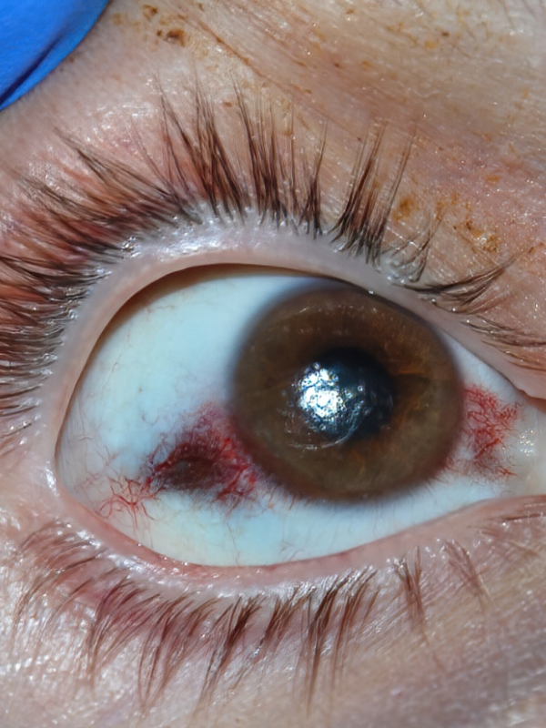
Symptoms of Pterygium
Symptoms vary based on the size and location of the pterygium and may include:
- Redness or irritation in the affected area
- Gritty sensation or discomfort in the eye
- Blurry or distorted vision, especially if the pterygium extends toward the center of the cornea
- Cosmetic concerns due to the visible appearance of the growth
When is Pterygium Surgery Necessary?
Surgery is considered when:
- The pterygium causes significant visual impairment.
- It causes persistent irritation or discomfort.
- It continues to grow or encroach on the central cornea.
- There are cosmetic concerns impacting a patient’s quality of life.
Treatment for Lacrimal Sac Conditions
1. Non-Surgical Treatments for Lacrimal Sac Conditions
For less severe cases, non-invasive therapies are employed:
- Antibiotic Eye Drops or Oral Medications:
If a lacrimal sac infection (dacryocystitis) is present, antibiotics can reduce infection and inflammation. - Warm Compresses:
Applying warm compresses to the affected area can relieve discomfort and improve drainage.
However, if these methods fail or the blockage persists, surgical intervention may be necessary.
2. Surgical Treatment for Lacrimal Sac Conditions
Surgical approaches aim to restore proper tear drainage by bypassing or addressing blockages or obstructions in the tear drainage system.
Types of Lacrimal Sac Surgery
- Dacryocystorhinostomy (DCR):
- The most effective surgical procedure to treat tear drainage obstructions.
- Creates a new connection between the lacrimal sac and the nasal cavity to bypass blocked tear ducts.
- Endoscopic DCR:
- A minimally invasive approach that uses an endoscope to access and repair the tear drainage system through the nasal cavity.
- Has a shorter recovery period compared to traditional surgery.
- External DCR:
- A traditional approach involving a small external incision near the corner of the eye.
- Suitable for patients who may not be candidates for the endoscopic approach.
- Tear Duct Stenting:
- Temporary or permanent stents are inserted into the lacrimal system to maintain proper drainage and prevent further obstruction.
Types of Lacrimal Sac Surgery
Dacryocystorhinostomy (DCR):
- The most common surgical procedure for lacrimal sac obstruction.
- A connection is created between the lacrimal sac and the nasal cavity to bypass the blocked tear duct.
Endoscopic DCR:
- A minimally invasive approach performed through the nose using an endoscope.
- Reduces recovery time compared to traditional external DCR.
External DCR:
- A traditional surgical method performed through a small incision near the nose to create a new tear drainage pathway.
Tear Duct Stenting:
- Temporary or permanent stents may be inserted to maintain patency and promote healing in cases of partial blockages.
Recovery and Aftercare
After lacrimal sac surgery, follow these steps to promote healing:
- Use prescribed antibiotics and anti-inflammatory eye drops: To prevent infection and inflammation.
- Manage nasal congestion: If you had endoscopic DCR, follow advice on managing nasal symptoms.
- Attend follow-up appointments: Monitoring is essential to ensure proper healing and tear drainage function.
- Avoid strenuous activities: Rest during the initial recovery period.
- Maintain proper hygiene: Keep the surgical site clean to minimize complications.
Most patients recover without complications and experience relief from symptoms like chronic tearing and infections.